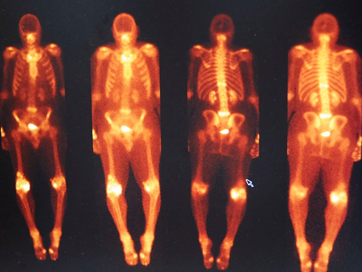
- During a nuclear bone scan, known as bone scintigraphy, patients may unexpectedly show high levels of DPD uptake (a cardiac radiotracer), indicating the presence of cardiac amyloidosis.
- Previously thought to be uncommon, recent advancements in diagnosis have revealed higher rates of CA than previously known.
- Now, new research has found that about 3% of individuals who undergo bone scans exhibit signs of CA, helping to gain further information on the occurrence and effects of this condition among the general population.
Cardiac amyloidosis (CA) causes the heart to thicken and become inflexible due to abnormal deposits of protein in place of healthy heart tissue.
The best way to treat this disease is to catch it early before it progresses. Otherwise, it can result in heart failure and even death.
A DPD scan is an imaging test that helps doctors diagnose CA. It uses a small amount of a radioactive substance called
During the test, these substances are injected into the body and then a special camera takes pictures of the heart. These images can show if the heart has abnormal deposits of the amyloid protein.
Amyloid deposits cause the heart muscles to become rigid, which makes it harder for the heart to pump blood throughout the body. Bone scintigraphy is therefore used to help doctors see how much amyloid has built up and how it is affecting the heart.
Since cardiac amyloidosis can lead to serious outcomes, it is crucial to accurately diagnose DPD uptake by the heart. People who display DPD uptake should be directed to a cardiologist for additional evaluation.
A new study, published in The Journal of Nuclear Medicine, is designed to determine the occurrence of cardiac amyloidosis in the general population through bone scans and to examine its impact on people with the condition.
Researchers recruited 11,527 individuals who received either cardiac or non-cardiac referrals and underwent DPD bone scintigraphy, generating a total…
Read the full article here


