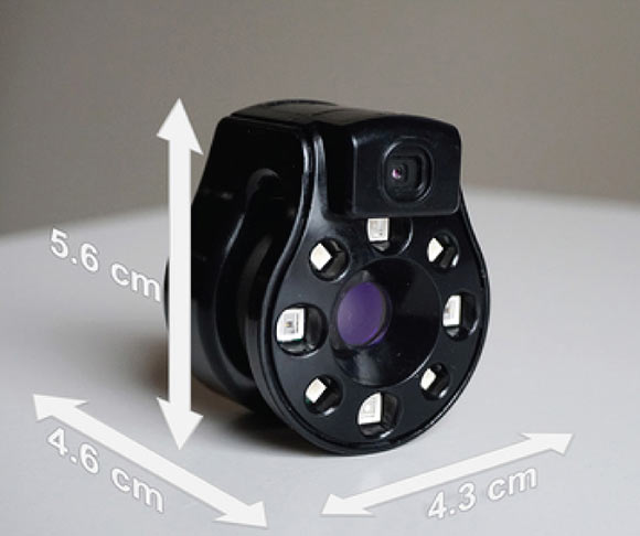
Named the Swift Ray 1, the real-life Star Trek ‘tricorder’ is a novel point-of-care hyperspectral imaging device that allows the acquisition of medical-grade images through a smartphone. Hyperspectral imaging acquires a multi-dimensional image dataset, called a hypercube, that provides diagnostic information about tissue physiology, morphology, and composition. The Swift Ray 1 device is equipped with near- and long-wave infrared sensors, violet light sources, and visible range LEDs, thereby allowing the simultaneous acquisition of visible light, infrared thermography, and bacterial fluorescence images as a hypercube. The device also integrates into the Swift Skin and Wound app, enabling precise wound area measurement, temperature quantification, and fluorescence area quantification.
“Wound care is one of today’s most expensive and overlooked threats to patients and our overall healthcare system,” said Dr. Robert Fraser, a researcher at Western University and Swift Medical Inc.
“Clinicians need better tools and data to best serve their patients who are unnecessarily suffering.”
The scientists developed a device called the Swift Ray 1 which can be attached to a smartphone and connected to the Swift Skin and Wound software.
This can take medical-grade photographs, infrared thermography images (which measure body heat), and bacterial fluorescence images (which reveal bacteria using violet light).
None of these images would be enough to identify infection alone.
Clinical inspection has low accuracy, as does thermography measuring heat changes caused by inflammation and infection.
Bacterial fluorescence can only look at the surface of a wound, which is naturally contaminated with bacteria, so additional methods are needed to differentiate between contamination and an infected wound.
“Research has demonstrated bacterial imaging helps guide clinicians’ work to remove nonviable tissue, yet it cannot identify infection by itself,” said Dr. Jose…
Read the full article here


