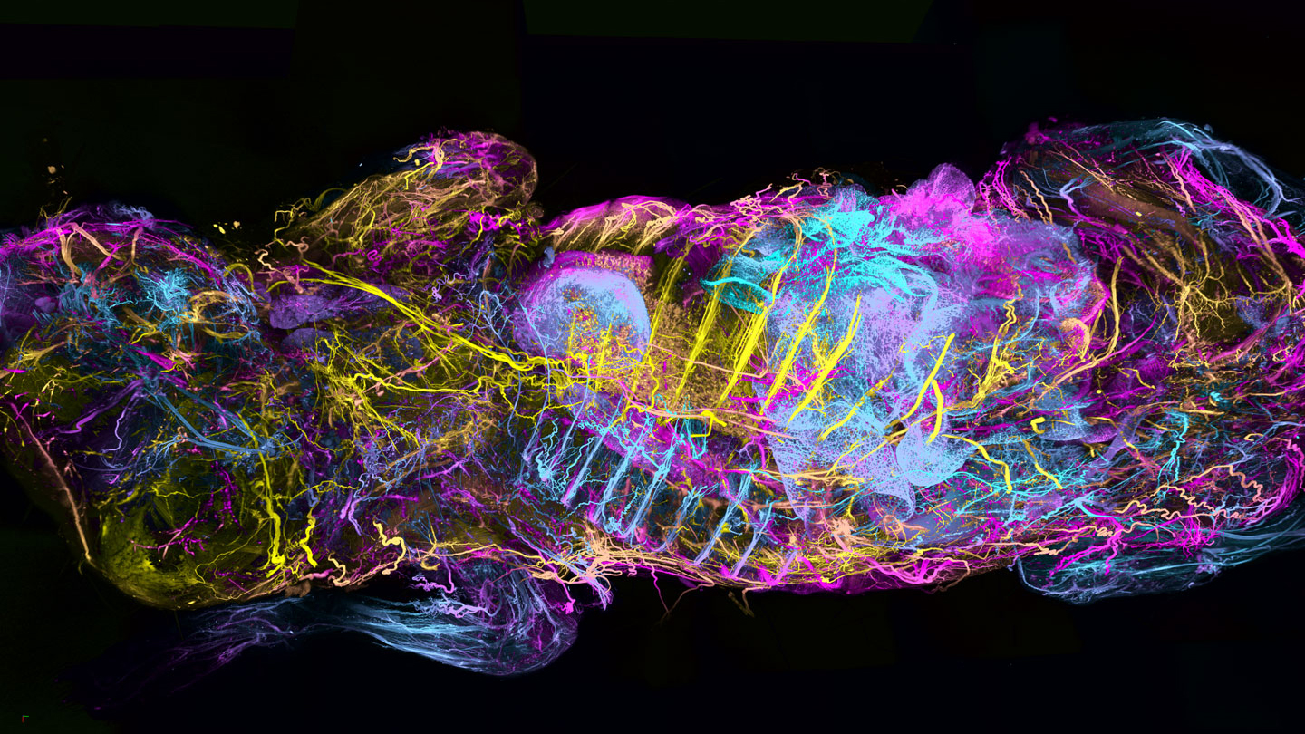
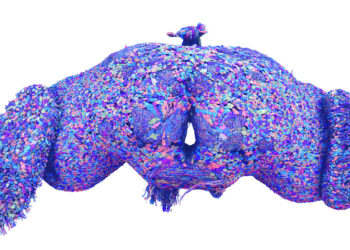
A mouse’s entire nervous system lights up in psychedelic hues. Clumps of immune cells attacking tumors give off a ghostly glow. The vessels that make up the body’s sewer system flare brightly. These and other images are shining a new light on the inner workings of mice. These glowing views are possible due to a new imaging technique. It makes imaging a whole body cheaper and faster.
Researchers described their colorful innovation July 10 in Nature Biotechnology.
To learn about the inner workings of a body, it helps to be able to see inside. See-through mice have been made before. But existing techniques to image the insides of whole animals can be expensive and time-consuming. Sometimes they don’t even hit the right target.
An improved technique starts by chemically removing cholesterol from the tissues of dead mice. That cholesterol is an essential part of cell membranes. Taking it out created spongelike holes in tissues — without destroying them. Those holes allowed better use of chemicals that can color, or label, structures of interest.
Targeted antibodies can move through the holes to reach every corner of the body. They bind to proteins of interest everywhere they reach. Under fluorescent light, they can make targeted parts of the body glow.
The technique gives scientists an amazingly thorough peek under mouse skin.
Called wildDISCO, it can be used to create atlases of the body’s interior. The process is a bit like Google Maps for the body, says Ali Ertürk. He’s a neuroscientist who led the work at Helmholtz Munich, in Germany. In place of cars driving around to record every street, this mapping system uses antibodies. They act as streetlamps to light up cellular landmarks.
Ertürk’s group first pumped cholesterol-removing chemicals into the hearts and blood vessels of dead mice. This allows liquids to flow through…
Read the full article here

