An eye full of cellular stars is a stunning example of the beauty that exists in nature’s smallest sizes.
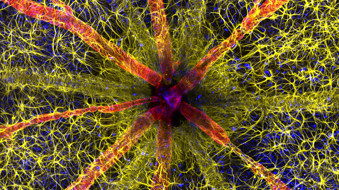
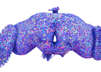
A glimpse of the back of a rat’s eye, and the immune cells that keep it healthy, won first place in the 2023 Nikon Small World photomicrography competition. The image, composed of multiple snapshots captured with a confocal microscope, was taken by neuroscientist Hassanain Qambari of the Lions Eye Institute’s Centre for Ophthalmology and Visual Science in Perth, Australia.
The photo is artificially colored to showcase the eye’s optic nerve — the black spot in the center — and surrounding structures in the retina, a layer of cells within the eye that captures light. A protein that helps blood vessels contract is shown in red and cell nuclei are blue. In yellow are astrocytes, a kind of immune cell that helps control inflammation in the retina.
The image is part of research that aims to uncover how diabetic retinopathy — a disease where high blood sugar damages retinal blood vessels — can alter the function and structure of the retina, Qambari says. When people are diagnosed, the disease is typically already in a late stage and the retina has sustained irreversible damage. Some people can go blind.
By pinpointing any changes that happen early on, researchers may be able to develop a drug to reverse those changes before the disease advances and causes damage.
The inner workings of the rat eye is one of 86 photos recognized in this year’s competition, the winners of which were announced October 17. Here are a few of our other favorites.
Fixing muscle
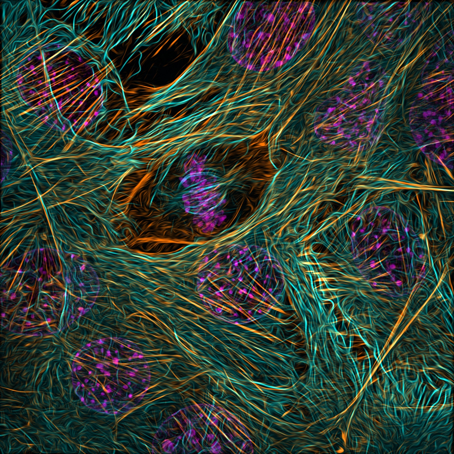
Some of these cells are in fix-it mode.
The photo, snapped by molecular physiologist Vaibhav Deshmukh, showcases cells called myoblasts that build muscle. These myoblasts are from mice and can be grown indefinitely in lab dishes. With dyes and antibodies, Deshmukh, then studying heart myoblasts as a graduate…
Read the full article here


