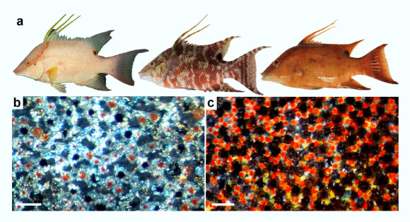
The hogfish (Lachnolaimus maximus), a common fish in the western Atlantic Ocean from North Carolina to Brazil, is known for its color-changing skin. The species can morph from white to mottled to reddish-brown in a matter of milliseconds to blend in with corals, sand or rocks.
Hogfish carry a gene for a light-sensitive protein called opsin that is activated in their skin, and that this gene is different from the opsin genes found in their eyes.
Other color-changing animals from octopuses to geckos have been found to make light-sensing opsins in their skin, too. But exactly how they use them to help change color is unclear.
“When we found it in hogfish, I said: Why have a light detector in the skin?” said Dr. Lori Schweikert, a biologist at the University of North Carolina Wilmington.
“One hypothesis is that light-sensing skin helps animals take in their surroundings.”
“But new findings suggest another possibility — that they could be using it to view themselves.”
In the study, Dr. Schweikert and colleagues took pieces of skin from different parts of the fish’s body and took pictures of them under a microscope.
Up close, a hogfish’s skin looks like a pointillist painting. Each dot of color is a specialized cell called a chromatophore containing granules of pigment that can be red, yellow or black.
It’s the movement of these pigment granules that changes the skin color. When the granules spread out across the cell, the color appears darker. When they cluster together into a tiny spot that’s hard to see, the cell becomes more transparent.
Next, the researchers used a technique called immunolabeling to locate the opsin proteins within the skin.
They found that in the hogfish, opsins aren’t produced in the color-changing chromatophore cells. Instead, the opsins reside in other cells directly beneath them.
Images taken with a transmission electron microscope revealed a previously unknown cell type, just below the chromatophores, packed with…
Read the full article here


